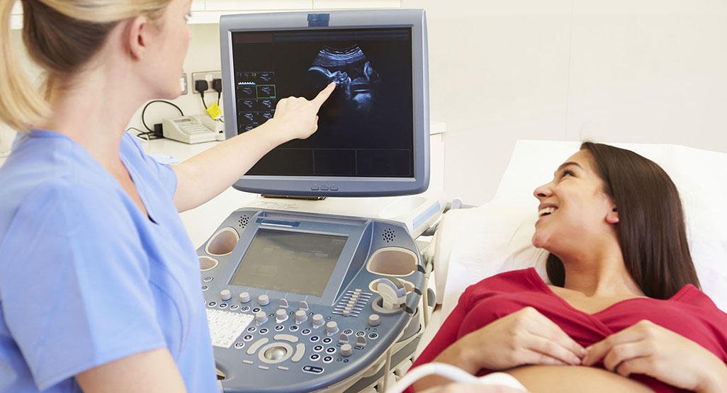
Like regular ultrasounds, 3D and 4D ultrasounds use sound waves to create an image of your baby in your womb.
3D/4D Ultrasound is a safe and widely used imaging technique. Ultrasound produces detailed images of the body in real-time using high-frequency sound waves which are produced by a special ultrasound probe. (Sonography) Ultrasound has no known harmful effects and can be used to image a variety of conditions including pregnancy, gallstones, and varicose veins.
3D Ultrasonography: 3D ultrasound is a medical ultrasound technique, often used in fetal, cardiac, trans-rectal, and intra-vascular applications. 3D ultrasound refers specifically to the volume rendering of ultrasound data and is also referred to as 4D (3-spatial dimensions plus 1-time dimension) when it involves a series of 3D volumes collected over time
4D Ultrasonography: 4D scans show moving 3D images of your baby, with time being the fourth dimension It’s natural to be excited by the prospect of your first scan.
When it is used?
3D/4D Ultrasound examinations can help to diagnose a variety of conditions and to assess organ damage following illness. Ultrasound is used to help physicians evaluate symptoms such as:
Pain
Swelling
Infection
Hematuria (blood in urine)
3D/4D Ultrasound is also used to:
Abdominal & Pregnancy Scan
Special Scans – Breast, Thyroid, Eye, Muscles & Joints
TVS/TRUS/Ovulation Study
3D/4D Ultrasound is a useful way of examining many of the body’s internal organs, including but not limited to the:
Heart and blood vessels, including the abdominal aorta and its major branches
Liver
Gallbladder
Spleen
Pancreas
Kidneys
Bladder
Uterus, ovaries, and unborn child (fetus) in pregnant patients